
Most of us have heard the statistic: about 1 in 8 women will develop invasive breast cancer over the course of their lifetime. It’s one of the deadliest cancers affecting women—in the United States, over 42,000 women die each year from the disease.1
Despite how common and consequential breast cancer is, surveys show that about 50% of women remain confused about the current guidance on screening.2 This confusion is not for lack of trying—the guidelines from organizations differ and have shifted multiple times, making it challenging for both patients and their doctors to determine the best option.
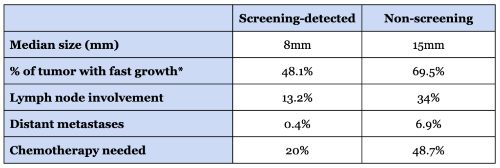
The good news is that breast cancer screening is extremely effective for reducing mortality: Women who screen regularly are between 25% and 42% less likely to die from breast cancer than women who do not.3,4 Cancers found through screening are more likely to be caught early—before they have spread—and tend to be smaller (about half the size of symptomatically-detected cancers; see Table 1) and easier to treat.5
Which raises the obvious question: if screening works this well, why are over 42,000 women still dying from breast cancer every year?
Part of the answer is biology. Some breast cancers are simply hard to catch. They grow quickly, spread early, or present in ways that routine screening is not well-designed to detect. By rough estimate, somewhere around 7–10% of breast cancers may fall into this category—cases that even a near-perfect screening system would struggle to intercept.
But there is another problem that is much more ordinary and more solvable: we are still under-screening.
That does not only mean that some women never get mammograms, though that is certainly part of it. Even among women who have been screened before, screening is often inconsistent. Roughly one-third of women over 40 have not had a mammogram in the past two years, and even among women aged 50–74—where the evidence for screening is least controversial—a substantial fraction are not up to date.6,7
Beyond just staying on schedule, there are also many women who are getting screened but with insufficient imaging for their risk profile. The clearest example of this is with breast MRI. Per the major screening guidelines, at least 9% of women meet the criteria for including breast MRI as part of their standard screening protocol. The actual utilization rate? 0.4%. This means fewer than 5% of women who should be getting more advanced testing are actually receiving the imaging the guidelines recommend.8 Moreover, many of these women are not even aware that they should be getting more advanced imaging.
It’s not that we lack the criteria for identifying high-risk women or that MRI is an unproven screening tool. This is not a scientific failure or a technology failure. It is an implementation failure: we already know how to identify many high-risk women and screen them more effectively, but in practice we are failing to match the right women to the right protocols.
So today, the goal is to help close that gap by moving away from one-size-fits-all screening and toward risk-matched screening. The goal is not just to tell you that screening is important—you already know that. The goal is to give you the necessary information for making more informed decisions about the specific screening protocol for you, based on your specific risk factors.
What the guidelines say
Before we get into personalizing screening, we need to start with the foundation: what do the published guidelines say?
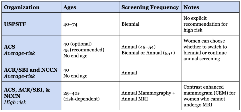
There are several organizations that put out guidance on breast cancer screening, including the American Cancer Society, the National Comprehensive Cancer Network, the American College of Radiology, and the US Preventive Services Task Force (USPSTF). Each differs on when to start and how often to screen—we’ve summarized their guidance in Table 2—so it’s not surprising that so many women are confused about screening. The USPSTF, whose guidance tends to inform insurance coverage decisions, recommends mammography only every other year for average-risk women aged 40 to 74, with no explicit recommendation for high-risk women. Most of the other groups are a bit more aggressive with their recommendations. Taking them together, the guidance is:
- Every woman should have a formal risk assessment by about age 25. Not because everyone needs imaging at 25, but because that is when you want to know whether you are truly average risk or someone whose screening should start earlier and look different.
- For average-risk women, annual mammography starts at age 40.
- For high-risk women, MRI starts much earlier—often by age 25 or 30—with annual mammography added between 25 and 40 depending on the specific risk profile.
- Screening continues as long as the woman would be willing to pursue treatment if cancer were found. There is no upper age cutoff for women who are healthy and would treat cancer if it were found.
These population-level guidelines are a good starting point for many average-risk women, but they aren’t necessarily the finish line—the moment you move from populations to actual people, personalization matters.
Our research team spends hundreds of hours each month vetting studies and distilling dense literature to deliver evidence-informed insights on health and longevity. If you find value in our work, consider becoming a premium member and supporting our mission.


A framework for personalizing screening
So how should we think about personalizing these guidelines? We can break this down into a few questions:
First, what is my baseline risk?
Second, given that risk, how much false-positive burden am I willing to tolerate in exchange for potentially earlier detection?
And third, when should I screen and which imaging modality or modalities are most appropriate for me?
The goal here is not to outsmart the guidelines—in fact, several of the guidelines explicitly stratify the screening strategy by risk level. The aim is to decide if the baseline approach—annual mammography beginning at 40—is actually the best option for you.
Assessing your risk
Starting with the first question, how do you determine your risk?
Most women who develop breast cancer do not have one dramatic risk factor. In fact, the two biggest risk factors are ones we rarely even think of as risk factors at all. Age is the strongest predictor of risk at the population level—the median age at diagnosis is 62, and the vast majority of cancers occur after age 40. Women are also far more likely to develop breast cancer than men (the lifetime breast cancer risk for men is about 1 in 755, compared to just 1 in 8 for women).
Beyond age and sex, it is far more common to have several smaller factors that together increase lifetime risk. So what other factors do we need to consider?
Genetics
Pathogenic mutations in genes like BRCA1, BRCA2, PALB2, ATM, and CHEK2 are by far the most powerful (and best-known) breast cancer risk factors. A woman with a BRCA1 mutation, for example, may have a lifetime risk of 60–70%, compared to about 12% in the general population.9 These mutations also shift risk earlier in life and toward more aggressive subtypes.
But pathogenic mutations are rarer than people tend to assume. Only about 1 in 400 people in the general population carries a BRCA1 or BRCA2 mutation, though the rate is much higher in some groups (roughly 2 in 100 in those of Ashkenazi Jewish descent).10 In total, only 5–10% of breast cancers are caused by known inherited mutations.11
Family history
While family history can capture the effect of high-impact mutations, it also reflects lower-impact mutations, shared environmental exposures, and other inherited factors that no single genetic test currently covers.
Multiple first- or second-degree relatives with breast cancer—especially those diagnosed at younger ages—can substantially increase risk. Family histories that include male breast cancer, ovarian cancer, or pancreatic cancer can also be meaningful, as those can signal an underlying BRCA mutation.
Family history is the factor that most of us point to when trying to assess our own risk for disease, but keep in mind that the absence of a known family history does not automatically equate to low risk.
Ancestry
Some ancestries carry meaningfully different breast cancer profiles. Some ancestries are associated with higher rates of certain mutations, as mentioned above, but others can influence risk through tumor biology or other factors. For example, non-Hispanic White women have a higher overall cancer rate, but Black women in the US tend to be diagnosed younger and with more aggressive subtypes of breast cancer. The precise mechanism for many ancestry-related associations is not known, but it can impact screening decisions.
Prior chest radiation
Exposure to high-dose radiation—particularly when breast tissue is rapidly developing—substantially increases breast cancer risk. The classic example is radiation treatment for Hodgkin lymphoma in adolescence or early adulthood. Cumulative dose, age at exposure, and number of exposures all matter.
Note that this does not mean you should avoid an occasional diagnostic chest X-ray—the dose from a routine X-ray is a tiny fraction of the dose used in cancer treatment, and the evidence that occasional imaging meaningfully increases breast cancer risk is weak.
Breast density
Dense breast tissue does two things: it independently raises baseline cancer risk, and it makes mammograms harder to interpret because both dense tissue and tumors appear white on the image. So density is not just a risk factor—it creates a dual problem by both increasing baseline cancer risk and making cancers harder to detect on mammography.
About half of screening-aged women have dense breasts, and high density is much more common at younger ages: roughly 60% of women in their 40s, 40% in their 50s, 30% in their 60s, and 28% in their 70s have dense breasts.12 Density is also fairly heritable (60–70%), so if your mother or sister has dense breasts, you are more likely to as well.13
The catch is that you usually cannot know your own breast density until you have had imaging—and most women do not until age 40. The FDA now requires mammography facilities to report breast density to patients, but that information typically arrives well after initial screening decisions have been made. We’ll come back to why this is relevant soon.
Reproductive and hormonal factors
Lifetime hormonal exposure plays a role in breast cancer risk. Factors that increase risk include earlier onset of menstruation, later menopause, never having had a full-term pregnancy, having a first pregnancy after age 30, and not breastfeeding.14
No single one of these typically changes a screening plan, but together they can shift the overall picture—especially when several are present.
Modifiable factors
A handful of lifestyle factors clearly affect risk. Alcohol consumption (even just a few drinks per week), obesity, and physical inactivity are all associated with higher risk for breast cancer.15 None of these are sufficient on their own to change your screening protocol, but it’s worth being aware of them since they are factors you can actually do something about.
Estimating your overall risk
No one can accurately estimate their breast cancer risk by adding all of these factors up in their head, so this is where formal risk calculators come in. The most widely used is the Tyrer-Cuzick model, which combines family history, personal risk factors, and breast density to produce a 10-year and lifetime risk estimate. It is not perfect, but it is meaningfully better than guessing—and it is the basis for most “high risk” classifications used clinically.
If your lifetime risk comes back above 20%, you are generally classified as high risk and qualify for additional screening.16,17 I think every woman should complete a formal risk assessment—including a calculator like Tyrer-Cuzick—by about age 25, both to know where she stands and to flag any need for earlier or more intensive screening.
| Action Item #1: Complete a formal risk assessment, as early as possible. Online calculators like the Tyrer-Cuzick model are a great start, but also think through the other risk factors mentioned above to see if that moves the needle on your screening protocol. |
Once you have that risk picture in hand, the next question becomes: how much false-positive burden are you willing to tolerate in exchange for the chance to catch cancer earlier?
Assessing your tolerance for false positives
In an ideal world, screening would find every meaningful cancer without any false positives. But of course, no screening test is perfect. Typically, the higher we boost a test’s sensitivity—that is, the better it is at finding cancer—the higher the false positive rate. More sensitive screening may also increase detection of lesions that would never have become clinically meaningful during a woman’s lifetime, which is part of the ongoing debate around screening intensity.
For breast cancer screening, the physical risk of the test is relatively minimal. The real burden comes from the emotional and logistical tolls of follow-up testing after an abnormal result. Having to return to the imaging center, take time away from work or family, and worry that this may be the start of a life-changing diagnosis is not trivial.
When a callback for additional testing leads to an earlier cancer diagnosis, it is obviously worthwhile. But if you haven’t experienced a callback before, this can all feel rather abstract. It helps to take a look at the numbers—just how common are false positives?
In the US, about 10% of screening mammograms lead to a callback for additional imaging or a biopsy. However, of those callbacks, only about 5% end in a cancer diagnosis.18 In other words, for every 1,000 women screened, roughly 100 will be called back because something looked abnormal. But of those 100, about 95 will not actually have cancer.
Those numbers add up over time. More than half of women who screen annually for 10 years will experience at least one false-positive result.18 This is a common occurrence, and it’s something we must factor in when developing our screening protocols.
How you weigh that tradeoff depends a lot on your baseline risk.
If you are very high risk, the math is easier. The probability that any given abnormal finding represents an actual cancer is much higher, and accepting more false positives in exchange for higher sensitivity is straightforward to defend.
If you are truly average-risk, the calculation becomes more nuanced. The pre-test probability of a true positive is lower, and you must think carefully about how much extra testing you are willing to accept for what may be a smaller incremental benefit. This is not to say you should avoid screening, but as we discuss the various options and their recall rates, keep this in mind.
The most important thing to keep in mind here is that more imaging is not automatically better screening. The objective is not to utilize as many imaging modalities as possible, but to land on the strategy that is most likely to benefit someone with your risk profile, without taking on any unnecessary false positives.
| Action Item #2: Be honest with yourself about your tolerance for false positives. If you are comfortable with some additional callbacks, knowing the relative probability of a true positive, you may lean towards more sensitive imaging. If the prospect of follow-up testing weighs heavily on you, particularly if your baseline risk is average or below-average, that may be a legitimate reason to stick closely to the default guideline. |
Once you know your risk and have thought about your tolerance for additional testing, the remaining questions are the practical ones: when do you start, how often do you screen, and which imaging modality should you use?
Imaging modalities
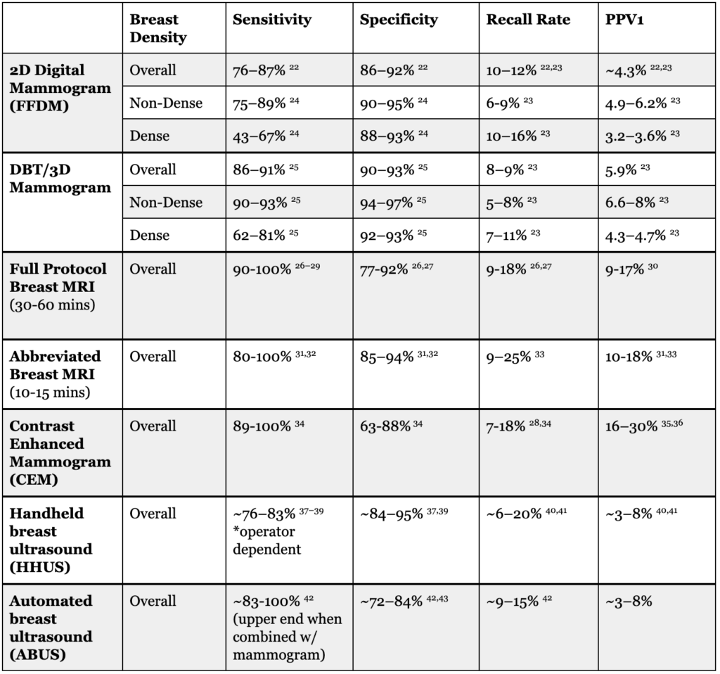
We’ll start with the imaging options. When we talk about screening for breast cancer, most of us immediately think of mammography. This is not unreasonable—mammography is the foundation for screening for virtually all women—but it is far from the only option available. You can see the detailed performance statistics for each option in Table 3.
Mammography
Mammograms use low-dose X-rays to look for breast cancer and are inexpensive and widely available. One important strength of mammography is that it is particularly good at detecting calcifications, such as those seen in ductal carcinoma in situ, or DCIS.
DCIS is sometimes called “stage 0” breast cancer. The abnormal cells are still confined to the ducts, rather than invading surrounding tissue. If left untreated, estimates suggest that somewhere between 25 and 60% of DCIS cases may eventually become invasive cancer.19 This wide range is a reflection of how much we still have to learn about DCIS, but the uncertainty is precisely why early detection is so critical.
That said, not all mammograms are the same.
In 2000, the FDA approved the first full-field digital mammography (FFDM) system, and this is what most of us think of when we hear “mammogram.” Standard digital mammography, sometimes called 2D mammography, is the technology that many research studies have historically used, and it remains the default at many imaging centers.
More recently, in 2011, digital breast tomosynthesis (DBT), commonly referred to as 3D mammography, was rolled out. DBT takes multiple images from different angles to create a more layered view of the breast, allowing for better cancer detection with lower recall rates, particularly for women with dense breasts.
Not every imaging center offers DBT, and sometimes there is still an added monetary cost. However, the data are very clear that DBT is a superior imaging modality and is worth prioritizing, particularly if you have dense breasts.
MRI
For women with higher baseline risk, MRI is the most sensitive option available. MRI uses magnetic fields and IV gadolinium-based contrast to detect abnormal blood flow or tissue behavior that mammography can miss. It is ideal for detecting very small invasive tumors and atypical cancers, though mammography remains better for some calcifications, so MRI is generally used in addition to, not instead of, mammography.
MRI comes in two options: full breast MRI and abbreviated breast MRI. The full breast MRI is typically recommended for very high-risk women, especially for initial screening, or as a diagnostic test after an abnormal result. The abbreviated breast MRI preserves nearly all of the sensitivity of the full protocol, but takes only 10 to 15 minutes compared to 30 to 60 minutes for the full exam.20 That makes it cheaper, faster, and more scalable—while still dramatically increasing cancer detection over mammography alone.
For women with extremely dense breasts, adding MRI after a negative mammogram cut the rate of interval cancers—meaning cancers found between screens—in half, from 5 per 1000 with mammography alone to 2.5 per 1000 with the addition of MRI.21 It’s worth noting that the recall rates for MRI can be higher than for mammography, though the likelihood of finding cancer (the positive predictive value 1, PPV1) is also higher. Part of this is likely due to study populations—most MRI studies are done in women already at higher risk—so it’s less clear how these rates may compare to mammography in average-risk women.
Regardless, all versions of MRI are more expensive than mammography, and can be harder to access. MRI also requires use of IV contrast, and is contraindicated for individuals with some conditions and medical devices (such as pacemakers or cochlear implants). But if the goal is maximal cancer detection, especially for women who are high-risk or have dense breasts, MRI’s sensitivity makes it an excellent option.
Contrast-enhanced mammography
The next best option is contrast-enhanced mammography (CEM). As its name suggests, this modality is essentially mammography plus IV iodine-based contrast. It gives more functional information than a standard mammogram and is a reasonable alternative when MRI is unavailable or contraindicated. This technology is fairly new—introduced in 2011—and is not yet widely available, but its higher sensitivity compared to standard mammography makes it a good option if it is available.
Ultrasound
The final option is ultrasound, which uses sound waves, rather than radiation or magnetic fields, to create images of breast tissue.
As with the other imaging modalities, ultrasound comes in different flavors: handheld, where a technician or radiologist moves the probe manually, and automated, where the machine acquires images more systematically. Handheld ultrasound is a good option once another imaging modality has flagged a potential issue, and is primarily useful for real-time evaluation of suspicious lesions and for guiding biopsies.
Compared to mammography or MRI, ultrasound (particularly handheld ultrasound) is more operator-dependent. It also carries a comparable recall rate to MRI, but with a lower likelihood that a recalled finding is actually cancer (PPV1). For routine screening, the benefit of ultrasound is more variable than the other modalities, but it does improve cancer detection over mammography alone and can be a good secondary imaging modality due to its lower cost and better access.
Deciding on imaging modalities
So how do we actually decide which imaging modality or modalities are best for our risk profile?
You can see a summary of the best use cases for each of the options discussed in Table 4. For virtually all women, mammography should remain the foundational screening modality. Depending on your risk, other modalities can be layered on top to improve detection.
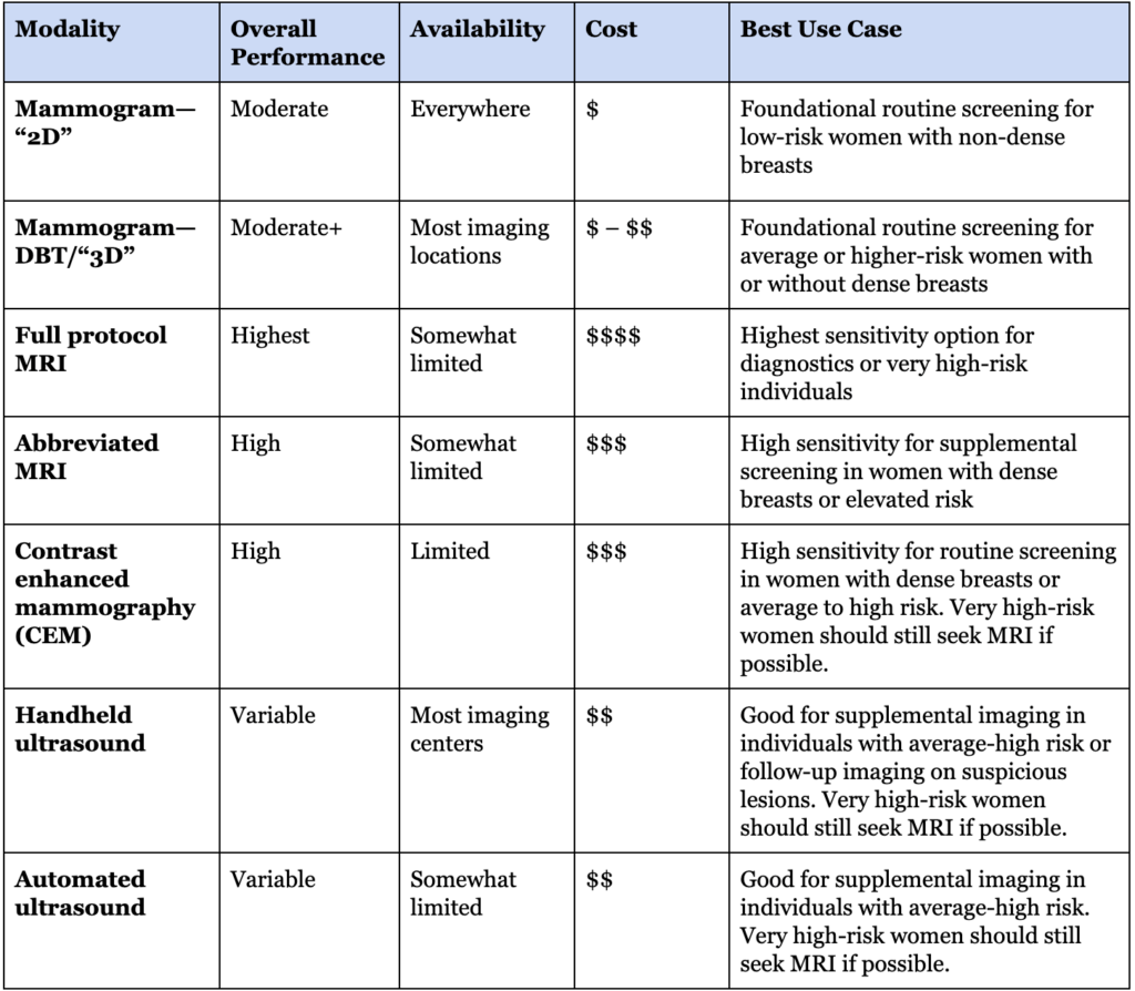
One important practical consideration is access. Not all imaging centers offer all modalities, and some centers are more experienced than others. Mammography is so common it can be performed well at many imaging centers. For low- or average-risk women with non-dense breasts, imaging is relatively straightforward and the particular center you attend may be less important. If you are pursuing more advanced imaging, such as MRI or CEM, or if you are at higher risk, have dense breasts, or have other considerations that may make imaging more complicated (such as breast implants), it may be worthwhile to go to a center with substantial expertise in breast imaging.
| Action Item #3: Think about which imaging modality or modalities make the most sense based on your risk profile and tolerance for false positives. Then identify options for imaging centers in your area that include those options and can provide the appropriate expertise for your situation. |
Screening intervals
Once you know which imaging tools make the most sense for you and have found a suitable imaging center, how often should you actually be screening?
This is another area where the guidelines diverge. The USPSTF recommends mammography every other year for average-risk women aged 40–74, while most radiology and cancer groups recommend annual screening starting at 40. The primary point of conflict between these groups is whether the goal is optimizing efficiency at the population level or maximizing cancer detection and survival odds for an individual woman.
We discussed the studies and context for why these groups came to different conclusions on our recent episode of The Drive, but to make a long story short: if the goal is maximizing the likelihood of early detection and reducing breast cancer mortality at the individual level, annual mammography is the strongest default position.
Things get murkier once you add in multiple imaging modalities. For women who undergo both mammography and MRI, many clinicians stagger them every six months—for example, a mammogram in January and an MRI in July. The logic is intuitive: it shortens the time between images that may detect a fast-growing cancer. The problem is that the evidence supporting this approach is actually quite limited. The same is true for ultrasound plus mammography; most studies performed both tests together annually, so there is very little direct evidence to guide how they should be spaced.
That means the most important priority is not whether the tests are perfectly staggered—it is whether they are actually getting done on schedule. For very high-risk women, especially those with genetic risk factors that may shift risk towards faster-growing cancers, alternating mammography and MRI every six months is a reasonable practical approach. For most other women using more than one modality, the timing can be guided by logistics, insurance coverage, and access. In many cases, simply getting both tests done at any point each year may matter more than whether they are separated by exactly six months.
| Action Item #4: Plan on annual mammography. If you are adding MRI or another modality, check your insurance coverage and work with your doctor to set a schedule that you can realistically maintain. The exact spacing matters less than being consistent and sticking to your plan. |
When to start screening
So at this point, you should know your baseline risk, and have a plan for when and where you are getting your selected imaging modalities. If you are 40 or older, you should already have a screening plan in place. But if you are below 40, when should you start screening?
As mentioned previously, most of the guidelines agree that age 40 is a reasonable time to start. And on average, the evidence for starting screening at 40 is solid. While the lifetime risk is about 1 in 8, only a small minority of breast cancers are diagnosed before age 40. Adding routine screening for everyone in their 30s is likely to expose many average-risk women to false positives and extra testing for a fairly small absolute yield.
The key here is that the women diagnosed before age 40 are not a random cross-section of the population: the majority of these cases occur in women with above-average risk. This is why the formal risk assessment in your 20s is so important. If you are identified as extremely high risk at 25, you may want to start screening in your 20s or early 30s. If you are average, or even below average risk, the default of 40 may be acceptable.
While the majority of the studies assessing screening women under 40 have looked at women with strong genetic risk factors, such as BRCA1 mutations, there is one study worth highlighting.44 In a large analysis of mammography data, researchers compared the cancer detection rate for women with or without common risk factors—dense breasts, a personal history of breast cancer, or a first-degree family history—across a variety of age groups.
Among women aged 35–39 with at least one risk factor, the cancer detection rate was 2.1 per 1,000 women screened. For average-risk women in the same age group—with no family history, no personal history, and non-dense breasts—the rate was just 0.59 per 1,000. That is about a three-and-a-half-fold difference driven by the presence of risk factors, not by age alone.
And here is where the comparison gets especially interesting. For average-risk women aged 40–44, the detection rate was only 0.71 per 1,000. In other words, women in their late 30s with risk factors were being diagnosed with cancer at roughly three times the rate of the average-risk women we already screen at 40.
Once you get to women 45 and older, incidence rises enough that detection rates exceed what we see in women under 40, regardless of risk factors. But below that threshold, risk factors may matter more than age.
This study also brings up another practical point. One of the risk factors was dense breast tissue—something that can only be definitely determined with imaging. About half of all screening-aged women have dense breasts, and density tends to decline with age, so while it is statistically more likely for younger women to have dense breast tissue, you can’t be certain until you start screening. So there is an argument to be made that a baseline mammogram in your 30s may be a way to further refine your risk profile. To be clear, there are no studies explicitly evaluating whether this improves outcomes—but for women who are already at a bit higher risk due to other factors, or who just want to have all possible information available when planning their screening, it’s not an unreasonable option for risk stratification. Keep in mind that your density will change over time, but if you have extremely dense breasts at 35, it’s unlikely you will drop down to the non-dense category by age 40.
With all this in mind, when should you start screening?
As with everything else, the answer depends on your risk. If you are low-risk or truly average-risk, beginning annual mammography at 40 is well-supported, though you could consider getting a baseline mammogram in your 30s to establish your approximate breast density. If you are above-average risk—for example, if you have a first-degree relative with breast cancer—there is a small amount of evidence that mammography in your 30s can be worthwhile. And if you are clearly very high risk—such as a known BRCA mutation carrier or a history of chest radiation—you’ll want to have a discussion with your healthcare team about a more aggressive screening protocol beginning in your 20s or 30s.
| Action Item #5: Plan to start screening by age 40 if you are average risk. Consider whether you want to add a baseline mammogram in your 30s to determine if you have dense breasts (bearing in mind that it will change over time). If you are high risk, discuss when to start with your doctor based on your specific risk factors. |
It’s also important to note that screening is for individuals without symptoms. Regardless of your age, if you experience any new breast symptoms—a lump, rash, pain, discharge, or anything else that isn’t normal for you—get it checked out. Screening isn’t perfect, so even a recent “normal” mammogram does not rule out the possibility of breast cancer if something seems off. This is especially important for inflammatory breast cancer, a rare but aggressive form of breast cancer that may not present as a classic lump. Instead, it can show up as rapid swelling, redness, warmth, changes in skin thickness or texture, or a change in breast shape. These symptoms can resemble infection or irritation, which is part of why diagnosis is sometimes delayed. Any new breast symptoms—even in women under 40, and even if they do not seem like cancer—are valid reasons to visit your primary care doctor or gynecologist for evaluation.
Putting it all together
So where does all of this leave us?
Breast cancer screening works. The tools are good, the evidence is strong, and screening allows many potentially deadly cancers to be found at an earlier, more treatable stage. We are never going to catch every cancer, but many breast cancer deaths are not inevitable—they happen because women were not screened, were not screened early enough, or were not screened with the right imaging.
The encouraging reality is that many of these failures are potentially preventable with better risk stratification and better adherence to appropriate screening protocols.
For many women, the right plan will still be the default: annual mammography beginning at age 40. But the only way to know whether that default actually fits you is to think through the questions we’ve covered here: What is my baseline risk? How much false-positive burden am I willing to accept? Which imaging modalities make the most sense for my situation? When should I start, and how often should I screen?
That is really the key point: screening should not be passive. It should be deliberate. The goal is not to get as much imaging as possible, nor to follow a population-level guideline without thinking about whether it truly applies to you. The goal is to build a screening plan that reflects the reality that breast cancer risk is not uniform, and therefore screening should not be uniform either.
If you take away only a few things, let them be these: know your risk early, choose the right imaging strategy for that risk, find an imaging center that can execute that plan well, and then follow through consistently. For most average-risk women, that means annual mammography beginning at 40. For women at higher risk, it may mean starting earlier and incorporating MRI or another modality. Either way, the objective is the same: risk-matched screening using the right tools, at the right intervals, for the right woman.
For a list of all previous weekly emails, click here.
References
1. SEER*Explorer. Accessed March 30, 2026. https://seer.cancer.gov/statistics-network/explorer/application.html
2. Adams S. Survey Finds Some Confusion Over Mammogram Guidelines. The Annenberg Public Policy Center of the University of Pennsylvania. June 30, 2025. Accessed May 12, 2026. https://www.annenbergpublicpolicycenter.org/survey-finds-some-confusion-over-mammogram-guidelines/
3. van Schoor G, Moss SM, Otten JDM, et al. Increasingly strong reduction in breast cancer mortality due to screening. Br J Cancer. 2011;104(6):910-914. doi:10.1038/bjc.2011.44
4. Dibden A, Offman J, Duffy SW, Gabe R. Worldwide review and meta-analysis of cohort studies measuring the effect of mammography screening programmes on incidence-based breast cancer mortality. Cancers (Basel). 2020;12(4):E976. doi:10.3390/cancers12040976
5. Starikov A, Askin G, Blackburn A, et al. Mode of detection matters: Differences in screen-detected versus symptomatic breast cancers. Clin Imaging. 2021;80:11-15. doi:10.1016/j.clinimag.2021.06.032
6. CDC. Challenges Women Face Limit Mammograms. Centers for Disease Control and Prevention. September 4, 2025. Accessed April 27, 2026. https://www.cdc.gov/vitalsigns/mammograms/index.html
7. Mammography. September 19, 2025. Accessed April 27, 2026. https://www.cdc.gov/nchs/fastats/mammography.htm
8. Lee MV, Aharon S, Kim K, et al. Recent trends in screening breast MRI. J Breast Imaging. 2022;4(1):39-47. doi:10.1093/jbi/wbab088
9. Cancer Risk for People with a BRCA1 Mutation. Cancer Risk for People with a BRCA1 Mutation. Accessed May 12, 2026. https://www.facingourrisk.org/info/hereditary-cancer-and-genetic-testing/hereditary-cancer-genes-and-risk/genes-by-name/brca1/cancer-risk
10. Levy R. What’s the Connection Between BRCA and Ashkenazi Jewish Ancestry? Dana-Farber Cancer Institute. June 26, 2023. Accessed May 12, 2026. https://blog.dana-farber.org/insight/2023/06/whats-the-connection-between-brca-and-ashkenazi-jewish-ancestry/
11. Apostolou P, Fostira F. Hereditary breast cancer: the era of new susceptibility genes. Biomed Res Int. 2013;2013:747318. doi:10.1155/2013/747318
12. Sprague BL, Gangnon RE, Burt V, et al. Prevalence of mammographically dense breasts in the United States. J Natl Cancer Inst. 2014;106(10):dju255. doi:10.1093/jnci/dju255
13. Boyd NF, Dite GS, Stone J, et al. Heritability of mammographic density, a risk factor for breast cancer. N Engl J Med. 2002;347(12):886-894. doi:10.1056/NEJMoa013390
14. Anothaisintawee T, Wiratkapun C, Lerdsitthichai P, et al. Risk factors of breast cancer: a systematic review and meta-analysis: A systematic review and meta-analysis. Asia Pac J Public Health. 2013;25(5):368-387. doi:10.1177/1010539513488795
15. Research and policy. World Cancer Research Fund. Accessed May 13, 2026. https://www.wcrf.org/diet-activity-and-cancer/
16. Accessed May 13, 2026. https://www.nccn.org/professionals/physician_gls/pdf/breast-screening.pdf
17. Saslow D, Boetes C, Burke W, et al. American cancer society guidelines for breast screening with MRI as an adjunct to mammography. Obstet Gynecol Surv. 2007;62(7):458-460. doi:10.1097/01.ogx.0000269073.50925.38
18. Mammogram False Positives Affect Future Screening Behavior. October 4, 2024. Accessed May 12, 2026. https://www.cancer.gov/news-events/cancer-currents-blog/2024/mammogram-false-positives-affect-future-screening
19. Wang J, Li B, Luo M, et al. Progression from ductal carcinoma in situ to invasive breast cancer: molecular features and clinical significance. Signal Transduct Target Ther. 2024;9(1):83. doi:10.1038/s41392-024-01779-3
20. van Grinsven SEL, Mann RM, Monninkhof EM, et al. Multireader diagnostic accuracy of abbreviated breast MRI for screening women with extremely dense breasts. Radiology. 2025;315(2):e241233. doi:10.1148/radiol.241233
21. Bakker MF, de Lange SV, Pijnappel RM, et al. Supplemental MRI screening for women with extremely dense breast tissue. N Engl J Med. 2019;381(22):2091-2102. doi:10.1056/NEJMoa1903986
22. Lehman CD, Arao RF, Sprague BL, et al. National performance benchmarks for modern screening digital mammography: Update from the Breast Cancer Surveillance Consortium. Radiology. 2017;283(1):49-58. doi:10.1148/radiol.2016161174
23. Conant EF, Talley MM, Parghi CR, et al. Mammographic screening in routine practice: Multisite study of digital breast tomosynthesis and digital mammography screenings. Radiology. 2023;307(3):e221571. doi:10.1148/radiol.221571
24. Payne NR, Hickman SE, Black R, Priest AN, Hudson S, Gilbert FJ. Breast density effect on the sensitivity of digital screening mammography in a UK cohort. Eur Radiol. 2025;35(1):177-187. doi:10.1007/s00330-024-10951-w
25. Kniss AS, Mercaldo S, Bahl M. Impact of breast density on screening performance metrics: An analysis of 301,400 screening digital breast tomosynthesis (DBT) examinations. Acad Radiol. 2026;0(0). doi:10.1016/j.acra.2025.12.042
26. Raikhlin A, Curpen B, Warner E, Betel C, Wright B, Jong R. Breast MRI as an adjunct to mammography for breast cancer screening in high-risk patients: retrospective review. AJR Am J Roentgenol. 2015;204(4):889-897. doi:10.2214/AJR.13.12264
27. Warner E. Screening BRCA1 and BRCA2 mutation carriers for breast cancer. Cancers (Basel). 2018;10(12):477. doi:10.3390/cancers10120477
28. Lawson MB, Partridge SC, Hippe DS, et al. Comparative performance of Contrast-enhanced mammography, abbreviated breast MRI, and standard breast MRI for breast cancer screening. Radiology. 2023;308(2):e230576. doi:10.1148/radiol.230576
29. Melnikow J, Fenton JJ, Whitlock EP, et al. Supplemental screening for breast cancer in women with dense breasts: A systematic review for the U.s. preventive services task force. Ann Intern Med. 2016;164(4):268-278. doi:10.7326/M15-1789
30. Lo G, Scaranelo AM, Aboras H, et al. Evaluation of the utility of screening mammography for high-risk women undergoing screening breast MR imaging. Radiology. 2017;285(1):36-43. doi:10.1148/radiol.2017161103
31. Kwon MR, Choi JS, Won H, et al. Breast cancer screening with abbreviated breast MRI: 3-year outcome analysis. Radiology. 2021;299(1):73-83. doi:10.1148/radiol.2021202927
32. Grimm LJ, Mango VL, Harvey JA, Plecha DM, Conant EF. Implementation of Abbreviated Breast MRI for Screening: AJR Expert Panel Narrative Review. American Journal of Roentgenology. 2022;218(2):202-212. doi:10.2214/AJR.21.26349
33. Gilbert FJ, Payne NR, Allajbeu I, et al. Comparison of supplemental breast cancer imaging techniques-interim results from the BRAID randomised controlled trial. Lancet. 2025;405(10493):1935-1944. doi:10.1016/S0140-6736(25)00582-3
34. Jochelson MS, Lobbes MBI. Contrast-enhanced mammography: State of the art. Radiology. 2021;299(1):36-48. doi:10.1148/radiol.2021201948
35. Sung JS, Lebron L, Keating D, et al. Performance of dual-energy contrast-enhanced digital mammography for screening women at increased risk of breast cancer. Radiology. 2019;293(1):81-88. doi:10.1148/radiol.2019182660
36. Patel BK, Carnahan MB, Northfelt D, et al. Prospective study of supplemental screening with contrast-enhanced mammography in women with elevated risk of breast cancer: Results of the prevalence round. J Clin Oncol. 2024;42(32):3826-3836. doi:10.1200/JCO.22.02819
37. Rehman H, Ahmad I, Rashid S, Mukhtar M, Khan AA, Khaliq H. Comparison of diagnostic accuracy of ultrasound and mammography in detecting breast cancer in radiographically dense breasts. Cureus. 2025;17(9):e92637. doi:10.7759/cureus.92637
38. Chen HL, Zhou JQ, Chen Q, Deng YC. Comparison of the sensitivity of mammography, ultrasound, magnetic resonance imaging and combinations of these imaging modalities for the detection of small (≤2 cm) breast cancer. Medicine (Baltimore). 2021;100(26):e26531. doi:10.1097/MD.0000000000026531
39. Sood R, Rositch AF, Shakoor D, et al. Ultrasound for breast cancer detection globally: A systematic review and meta-analysis. J Glob Oncol. 2019;5(5):1-17. doi:10.1200/JGO.19.00127
40. Brem RF, Lenihan MJ, Lieberman J, Torrente J. Screening breast ultrasound: past, present, and future. AJR Am J Roentgenol. 2015;204(2):234-240. doi:10.2214/AJR.13.12072
41. Berg WA, Blume JD, Cormack JB, et al. Combined screening with ultrasound and mammography vs mammography alone in women at elevated risk of breast cancer. JAMA. 2008;299(18):2151-2163. doi:10.1001/jama.299.18.2151
42. Choi WJ, Kim SH, Shin HJ, et al. Automated breast US as the primary screening test for breast cancer among East Asian women aged 40-49 years: a multicenter prospective study. Eur Radiol. 2021;31(10):7771-7782. doi:10.1007/s00330-021-07864-3
43. Gatta G, Cappabianca S, La Forgia D, et al. Second-generation 3D automated breast ultrasonography (prone ABUS) for dense breast cancer screening integrated to mammography: Effectiveness, performance and detection rates. J Pers Med. 2021;11(9):875. doi:10.3390/jpm11090875
44. Lee CS, Ashih H, Sengupta D, Sickles EA, Zuley M, Pisano E. Risk-based screening mammography for women aged <40: Outcomes from the National Mammography Database. J Am Coll Radiol. 2020;17(3):368-376. doi:10.1016/j.jacr.2019.08.033


